First, keep the image in the right place
MIT's October 19 account describes E(3)-Pose, a research model developed with Boston Children's Hospital and Inria to help account for fetal movement during MRI. The aim is to obtain images a radiologist can examine, not to replace that examination.
The practical problem is easy to picture. An MRI examination can build a volume from a sequence of thin image slices. If the object moves between slices, the sequence can miss areas or capture them at unintended angles. MIT presents movement-aware acquisition as a way to address that problem.
Following a turn rather than naming a condition
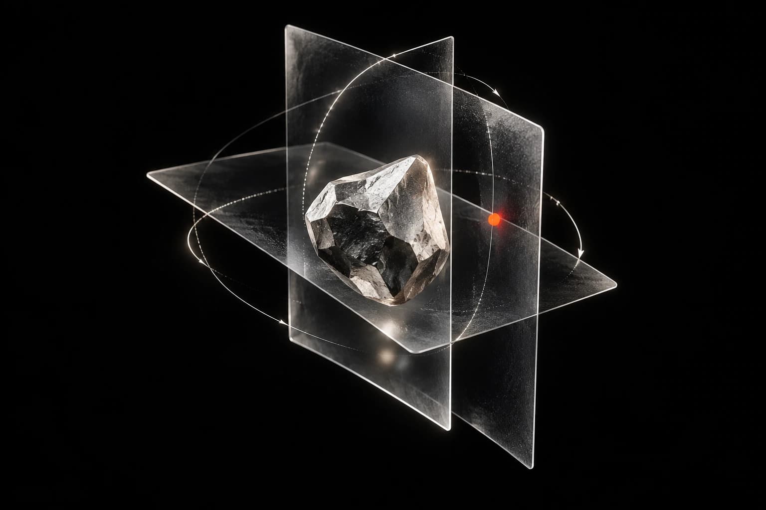
The paper describes using rapid, low-resolution three-dimensional images to estimate head position and orientation before subsequent slices. It builds rotations and approximate left-right symmetry into the model's design.
This idea is called equivariance: when an input rotates, the estimated orientation should change consistently with it. The purpose is a stable spatial reference, not a medical finding.
A simple distinction helps. Knowing which way an object faces can guide where the next image is taken. It does not say whether the object contains a particular abnormality. These tasks need different reference answers and different evaluation.
What the evidence supports
The arXiv record shows the work was first submitted in December 2025 and last revised on June 25, 2026. The latest MIT article is a new account of existing research, not evidence that the study itself began this week.
The paper evaluates position estimates on research and clinical image datasets. Its navigator dataset contains repeated volumes from nine volunteers. Repeated images provide many test inputs, but do not turn that group into an equally large number of independent participants.
The authors also report simulations of adaptive slice placement. Their appendix describes scanner implementation, while leaving larger-cohort evaluation of diagnostic usefulness to future work. The paper notes limitations around major asymmetries and uncertainty estimation.
How to read the next claim
MIT's account describes potential benefits for image quality and patient experience. Those are goals to test, not proof that this model reduces missed diagnoses, repeat visits or complications. This desk has not run the software or independently reproduced its results.
A useful evidence checklist has three parts: does the model locate the object reliably; does the complete scanner workflow produce useful images; and does that change clinical decisions or outcomes? Success at one stage is a reason to test the next, not to assume its answer.
For readers, the takeaway is narrow but useful. AI can contribute to how medical data are acquired, even when it does not interpret a diagnosis. That contribution still needs to be judged in the complete workflow where the images will be used.
Sources
- MIT News: symmetry and fetal MRIOctober 19 institutional account of E(3)-Pose and potential imaging benefits. Researcher expectations not treated as proven diagnostic or patient outcomes.
- arXiv: E(3)-Pose version historyPrimary version record: first submitted December 4, 2025; latest v8 June 25, 2026. Current institutional news is not a newly released study.
- E(3)-Pose full paper and appendices, v8Primary technical methods, datasets, simulations, implementation and limitations reviewed. Repeated navigator volumes from nine volunteers; diagnostic evaluation left to future work. No independent reproduction or clinical-outcome claim.